Reseña o resumen
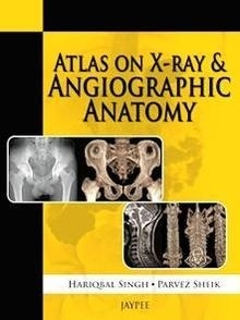
This atlas presents trainees with numerous X-ray and
angiographic images to gain a thorough understanding of
normal radiographic anatomy in order to make an accurate
diagnosis of underlying pathology.
Presented in an easy to read format, the book covers
radiological procedures, ossification centres, X-ray
production, digital subtraction angiography, and computed
and digital radiography, in the different anatomical sections
of the body.
This practical guide includes nearly 240 clearly labelled
images, illustrations and tables, with detailed descriptions,
to assist learning.
Key points
Atlas of X-ray and angiographic images to help trainees
understand normal radiographic anatomy and diagnose
underlying pathology
Easy to read format
Covers different imaging techniques for all areas of the
body
Includes nearly 240 images, illustrations and tables with
detailed descriptions.
Table of Contents
1. Skull
2. Spine
3. Chest
4. Abdomen
5. Upper Limb
6. Lower Limb
7. Angiograms
8. Radiological Procedures
9. Ossification Centers
10. Production of X-rays
11. Digital Subtraction Angiography
12. Computed and Digital Radiography
13. Picture Archiving and Communications System
14. CT Contrast Media