Reseña o resumen
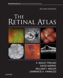
*2011 BMA Awards - Highly Commended in Internal Medicine *Dr. Lawrence A. Yannuzzi brings together the most complete retinal atlas ever. Over 5,000 illustrations of the latest imaging and research findings essential for effective diagnosis of retinal disorders populate The Retinal Atlas. A unique page layout consisting of optimally positioned panoramic images, magnified photos, and histopathological specimens illustrate key manifestations, giving you the best visual display of each disease. In addition, composite images using different retinal imaging modalities, including the latest in optical coherence tomography (OCT), fluorescein angiography, indocyanine green (ICG), and fundus autofluorescence display how a disease appears in each imaging modality, allowing you to compare imaging methods and gain a better understanding of each disorder. The Atlas is the ideal resource for all retinal specialists, comprehensive ophthalmologists, and other eye care personnel. The Expert Consult functionality gives you easy access to the full text online, as well as a downloadable image library at expertconsult.com.
Key Features
Includes full-text online access to the complete contents of the book, a downloadable image library, and links to Medline at expertconsult.com.
Features complete, comprehensive coverage of all vitreous, retina, and macula diseases, assimilating old and new photos for effective diagnosis at early and later stages of each disorder.
Covers all new imaging methods used to present and illustrate retinal diseases, including the latest on ophthalmic coherence tomography, indocyanine green angiography, fluorescein angiography, and fundus autofluorescence, keeping you up to date with new, developing, and cutting edge imaging techniques to match evolving diagnosis and treatment methods.
Incorporates arrows and guides into the images that point to key lesions for a more accurate identification of disorders.
Provides a unique page design using composite layouts that incorporate various forms of disease presentation, including high-power views and the latest panoramic photos, offering an enhanced understanding of the full spectrum of disorders.
Offers concise coverage of key histopathology findings, providing an improved understanding of the clinico-pathological relationships and selected references for additional readings.
Presents a select team of industry experts, all of whom are true international leaders in their sub-specialty areas, and have assisted in contributing to the diverse library of common and rare case photos.
Chapter 1: Normal
Chapter 2: Hereditary chorioretinal dystrophies
Chapter 3: Pediatric retina
Chapter 4: Inflammation
Chapter 5: Infection
Chapter 6: Retinal vascular disease
Chapter 7: Degeneration
Chapter 8: Oncology
Chapter 9: Macular fibrosis, pucker, cysts, holes, folds, and edema
Chapter 10: Non-rhegmatogenous retinal detachment
Chapter 11: Peripheral retinal degenerations and rhegmatogenous retinal detachment
Chapter 12: Traumatic chorioretinopathy
Chapter 13: Complications of ocular surgery
Chapter 14: Chorioretinal toxicities
Chapter 15: Congenital anomalies of the optic nerve